Three-dimensional ultrasound enables real-time volumetric visualization of anatomical structures. Unlike traditional 2D ultrasound, 3D imaging reduces reliance on precise probe orientation, potentially making ultrasound more accessible to clinicians with varying levels of experience and improving automated measurements and post-exam analysis. However, achieving both high volume rates and high image quality remains a significant challenge. While 3D diverging waves can provide high volume rates, they suffer from limited tissue harmonic generation and increased multipath effects, which degrade image quality. One compromise is to retain focus in elevation while leveraging unfocused diverging waves in the lateral direction to reduce the number of transmissions per elevation plane. Reaching the volume rates achieved by full 3D diverging waves, however, requires dramatically undersampling the number of elevation planes. Subsequently, to render the full volume, simple interpolation techniques are applied. This paper introduces a novel approach to 3D ultrasound reconstruction from a reduced set of elevation planes by employing diffusion models (DMs) to achieve increased spatial and temporal resolution. We compare both traditional and supervised deep learning-based interpolation methods on a 3D cardiac ultrasound dataset. Our results show that DM-based reconstruction consistently outperforms the baselines in image quality and downstream task performance. Additionally, we accelerate inference by leveraging the temporal consistency inherent to ultrasound sequences. Finally, we explore the robustness of the proposed method by exploiting the probabilistic nature of diffusion posterior sampling to quantify reconstruction uncertainty and demonstrate improved recall on out-of-distribution data with synthetic anomalies under strong subsampling.
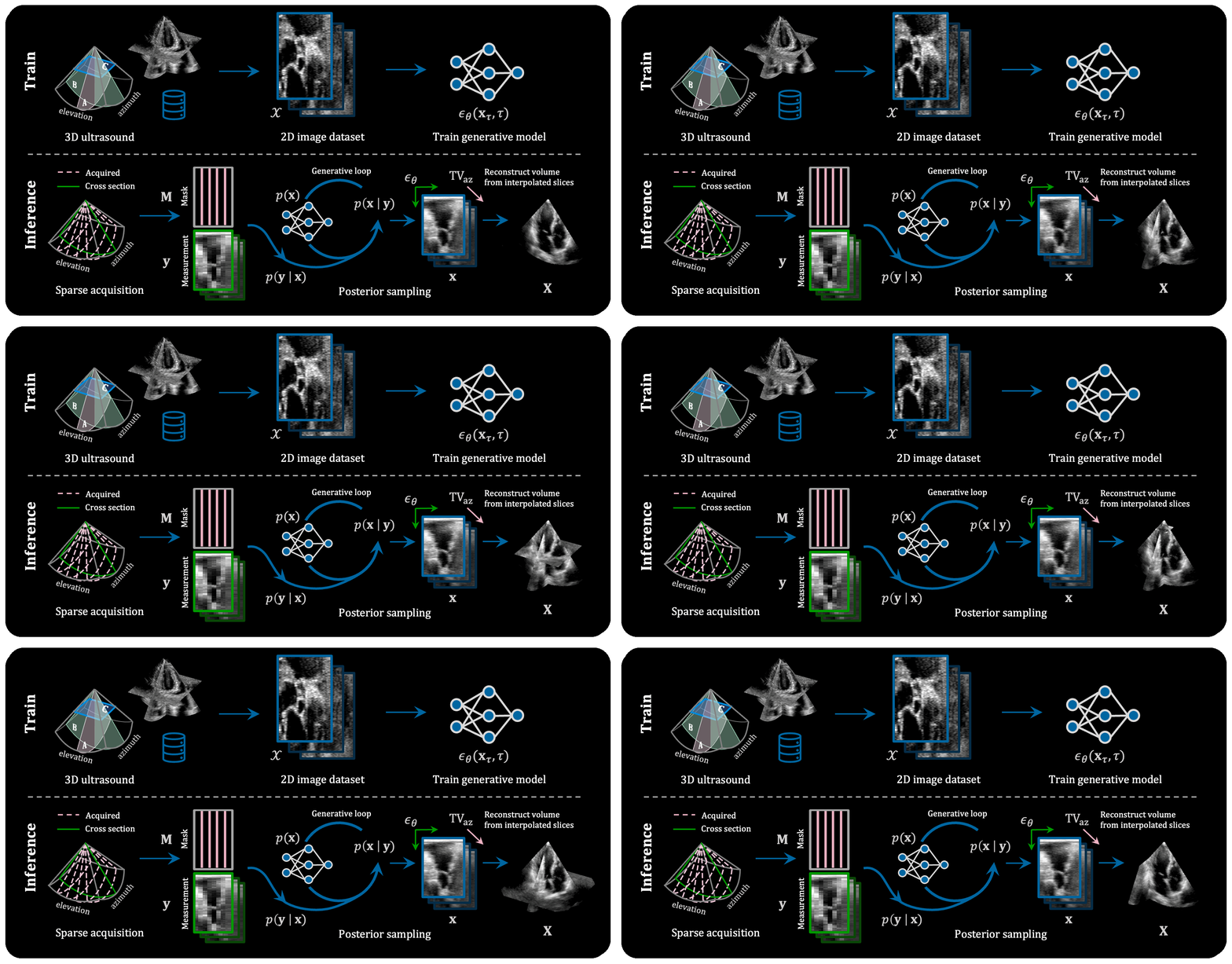
Overview of the training and inference pipeline for diffusion-based interpolation of 3D ultrasound volumes. A 2D score-based prior is learned from fully sampled elevation (B-plane) slices extracted from the training dataset. At inference time, this generative prior guides the posterior sampling process to reconstruct from subsampled B-planes.
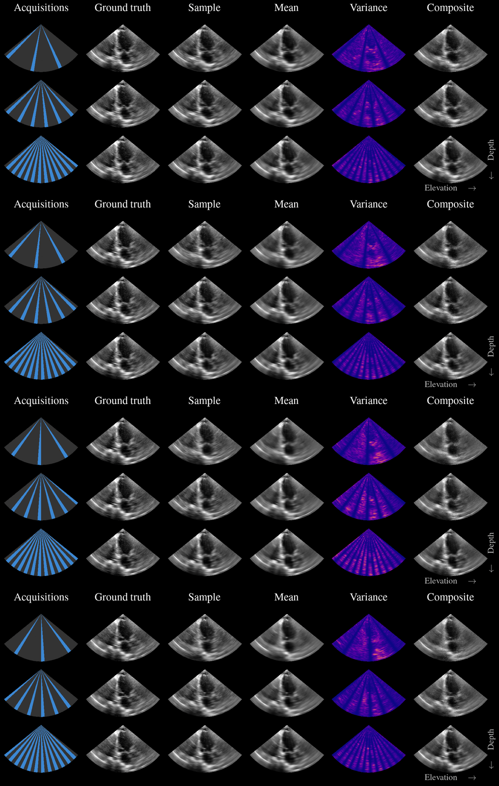
Many interpolation methods provide only a single point-estimate reconstruction, failing to represent the uncertainty inherent to the inverse problem. Given the probabilistic nature of diffusion posterior sampling, however, our method can quantify the uncertainty present in its reconstructions. Quantifying the uncertainty can help mitigate hallucinations, in which a model generates one of many plausible anatomies that may be convincing but not truthful. Or, more critically, it overlooks an anatomical feature that is, in fact, present. In our paper, we describe two methods to quantify and communicate this uncertainty. We build on the well-established idea of combining samples from the posterior distribution. Our contribution lies in adapting this principle to 3D ultrasound reconstruction and proposing visualization strategies that convey the uncertainty in an interpretable manner to the operator.
Compare different 3D ultrasound reconstruction methods side by side using the slider below. All images are reconstructed with acceleration rate r=3.
The official implementation is available at https://github.com/tue-bmd/3d-ultrasound-diffusion. It is built on zea (docs), a toolbox for cognitive ultrasound imaging, with the JAX backend. zea provides the generative modeling infrastructure, including posterior sampling, as well as the ultrasound signal processing pipeline and data handling (including streaming datasets from the Hugging Face Hub). For a simplified 2D version of this approach, i.e. diffusion posterior sampling of a single subsampled image, see the zea diffusion model example.
@article{stevens2025highvolumerate3d,
title={High Volume Rate 3D Ultrasound Reconstruction with Diffusion Models},
author={Stevens, Tristan S. W. and Nolan, Oisín and Somphone, Oudom and Robert,
Jean-Luc and van Sloun, Ruud J. G.},
year={2025},
journal={IEEE Transactions on Medical Imaging},
doi={10.1109/TMI.2025.3645849}
}